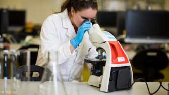
The Mark Wainwright Analytical Centre
A central facility supporting ESSRC research
A central facility supporting ESSRC research

The Mark Wainwright Analytical Centre (MWAC) is a network of centralised cutting-edge facilities and expert staff that are open to the entire UNSW research community and beyond. The MWAC currently comprises the following units:
For more information about the MWAC or its constituent units, see the Mark Wainwright Analytical Centre (MWAC).
Many MWAC staff have research interests aligned with the ESSRC research centre, and some have been recognised as affiliates of ESSRC. ESSRC affiliates are currently based in the Bioanalytical Mass Spectrometry Facility (BMSF) and the Electron Microscope Unit (EMU).
Bioanalytical Mass Spectrometry Facility UNSW brings together advanced mass spectrometric equipment and allied technologies, software, bioinformatics, and staff expertise in all aspects of protein, metabolite and lipid, macro and micro molecule analysis. Analytical applications focus on the identification and characterisation of molecules from biological and archaeological contexts. The BMSF can provide students, researchers, and academics the guidance, training and method development specific to each ESSRC project. Collaboration or fee-for-service is invited.
ESSR research projects undertaken by BMSF staff highlight the specialised capabilities possible.
Paleo proteomics/lipidomics/metabolomics is the scientific study of ancient molecules. This area of research is gaining interest to support the various theories of human migration through ‘deep-time’. It provides a new view of the past that takes advantage of the sometimes exceptional preservation above and beyond the fragility of DNA, particularly in climates that quickly degrade DNA, the very regions with the greatest biodiversity.
https://analyticalsciencejournals.onlinelibrary.wiley.com/doi/10.1002/pmic.201800341
https://www.mdpi.com/1422-0067/21/17/6422
Stable isotope analysis (SIA) measures the ratios of the stable isotopes of Carbon (12/13C), Oxygen (16/18O) and Nitrogen (14/15N) as a gas, which is compared to standard reference gases. The BMSF assays both inorganic and organic samples for paleoclimate, paleontological, environmental, and ecosystem studies.
Inorganic SIA applies very small amounts of phosphoric acid to calcite (speleothem, bone apatite, coral, shells, or sediment) samples. This liberates CO2, which is then analysed by the mass spectrometer to give δ13C and δ18O. The study of the carbon and oxygen isotopes in speleothems and coral provide a past climate proxy, and in bone help determine the environments humans or other animals lived and what they ate (diet). In shells, they can provide a proxy of ocean temperature for marine forensic science.
Organic SIA samples undergo combustion in an oxygen atmosphere and are converted into simple gases (such as CO2 and N2). The evolved gases are ionised and separated before being detected by the mass spectrometer. Measurement of the ratio of the ions can provide information about the biological, chemical or physical processes the material has undergone. This has a wide range of applications such as predation, food webs, migration patterns and ecosystem management.
https://www.sciencedirect.com/science/article/abs/pii/S0016703719307641?via%3Dihub
https://www.sciencedirect.com/science/article/abs/pii/S2352409X18306163
https://link.springer.com/article/10.1007/s00227-021-03822-1
Lewis Adler
Senior Technical Officer
Bioanalytical Mass Spectrometry Facility
Mark Wainwright Analytical Centre
Lab B50, Chemical Sciences Building F10
Ph:+61 2 93857739
Web: http://www.analytical.unsw.edu.au/facilities/bmsf
Kiel You-Tube from UNSW TV: https://youtu.be/K3_NLVDoGVg
Stalagmites and stalactites are a type of speleothem, a calcium carbonate deposit formed in limestone caves. These deposits form from drip water that is supersaturated with respect to calcite or aragonite due to the dissolution of carbonate bedrock between the surface and the cave. It’s well known that trace elements (e.g., Mg, S, P, Sr, Ba, Fe, Zn) incorporated into the calcareous stalagmites and stalactites provides information on past environmental change.
Helen Wang, Micheline Campbell, Andy Baker and ANSTO colleagues examined the annual variations of trace elements, e.g., Mg, P, S and Sr along stalactite laminae using M4 Tornado micro-XRF to understand the past climate change in Yanchep, Western Australia over the last few hundred years. Use of the micro-XRF enabled the research team to non-destructively measure annual variations in strontium concentration in this very fragile sample. The strontium data was used to build a chronology for the stalactite deposition – high strontium occurs in the calcite deposited in the dry season and low strontium in the wet season. The team will use this chronology and analyse other trace elements, to reconstruct past fire history and climate variability, funded by an ARC Discovery Project.
Strontium 20 mm line scan and mapping analysis of a ‘soda-straw stalactite’ sample (top). Mapping area of 20 mm x 0.3 mm is boxed in green. Bottom figure shows Sr/Ca five point centred moving average. Red star shows Sr/Ca peaks used to build a chronology for the stalactite deposition.
The Chronos Facility at UNSW is a radiocarbon dating laboratory that employs an Ionplus Mini CArbon DAting System (MICADAS) paired with an Elemental Analyzer and Automated Graphitization System (AGE3) as well as a Carbonate Handling System (CHS2). Traditionally 14C-AMS dating can be costly, time-consuming and, depending on the method, require large sample volumes. The advantage of a MICADAS system is that high-throughput of samples is possible using relatively small sample sizes – for example, 10mg of wood is sufficient to produce a full-sized graphite target for analysis, whereas at other labs, 100mg of wood is the standard starting volume. The ability for high sample throughput at a lower cost is of great advantage to ESSRC researchers as there are several studies currently being undertaken that require larger quantities of radiocarbon measurements than would have been possible through commercial dating facilities.
At Chronos, sample preparation can be done in house with pretreatment protocols and matrix matched backgrounds available for wood, carbonates, charcoal, peat, pollen, and bone. We also welcome samples pretreated from other facilities where a description of the pretreatment processes is well documented and accompanies the samples.
Heather Haines, Chris Turney, Jonathan Palmer, Zoë Thomas, and Chris Marjo are undertaking a large-scale project to identify Solar Energetic Particle (SEP) events of differing magnitudes in Southern Hemisphere tree-rings. Along with identifying these events the team is looking to better understand the seasonality of the larger SEPs. For this project individual tree-rings have been subsampled into sizes as small as 5mg of raw wood which has provided reliable C14 measurements on the MICADAS – something not possible with many other AMS machines.
Zoë Thomas is leading work creating high-resolution chronologies of sediment sequences from the southern mid-high latitudes. Developing accurate age-depth models from peat sequences is difficult due to the commonality of outliers caused by issues such as root penetration and sediment in-wash. However, the Chronos protocols permit careful consideration when selecting materials to date with the ability to date smaller material such as macrofossils when available. The results provide highly precise chronologies with which to test the timing of global atmospheric circulation changes.
Heather Haines and colleagues are undertaking radiocarbon dating on tropical Australian tree species which are known to present issues when using traditional dendrochronological dating methods due to anomalous ring boundaries. The use of radiocarbon dating allows for an understanding of the nature of a species growth and permits the development of robust annual tree-ring records that can be used to reconstruct past climate in parts of Australia where long-term instrumental records do not exist and other paleoclimate proxies are scarce.
Prof. Chris Turney
Director of the Earth and Sustainability Science Research Centre (ESSRC)
Director of the Chronos 14Carbon-Cycle Facility
Email: c.turney@unsw.edu.au
The facilities and expertise of the EMU are targeted toward imaging and spatially-resolved microanalysis, from the millimetre to the nanometre scale. The EMU provides ESSRC, UNSW, external and commercial researchers to high-end microscopy techniques such as:
For a comprehensive and current list of EMU microscopy and specimen preparation facilities, see http://www.analytical.unsw.edu.au/facilities/emu/instruments.
The EMU is the largest unit of the MWAC, and provides training and research support to internal researchers, as well as those from other domestic and international universities, government agencies, and commercial entities. EMU staff also collaborate on research projects, from concept development through presentation and publication.
In addition to the wide range of advanced microscopy techniques provided and supported in-house by the EMU, the EMU’s participation Microscopy Australia (MA) provides users access to additional world-class techniques and expertise based at partner institutions around Australia, such as nano-SIMS (UWA) and Atom Probe Microscopy (University of Sydney). In partnership with other MA members, the EMU has helped to develop MyScope, an online tool now used across the world to teach theory and operation of a variety of microscopy and microanalysis techniques to students and researchers.
Microscopy Australia: micro.org.au
MyScope Online Microscopy Training and Simulators: myscope.training
Some recent research conducted at the EMU by ESSRC staff and affiliates:
Tara Djokic, Martin van Kranendonk and colleagues examined ca. 3.45 Ga stromatolites using techniques including SEM-EDS to extend the known geological record of inhabited terrestrial hot springs on Earth by around 3 billion years.
https://www.nature.com/articles/ncomms15263
Chris Turney, Zoë Thomas, Karen Privat and colleagues used SEM imaging to examine the morphology of sediment grains to aid reconstruction of ancient atmospheric circulation regimes over southern NZ.
http://www.sciencedirect.com/science/article/pii/S0277379116307144
Karen Privat and colleagues examined the morphology of early colonial Peruvian pottery sherds to reconstruct firing techniques and technologies employed by the indigenous population in the period immediately following the Spanish invasion.
http://www.sciencedirect.com/science/article/pii/S0305440317300547
Sue Hand used SEM facilities at the EMU to characterise the dentition of a new archaic bat from the Eocene.
Samples prepared for electron probe microanalysis (EPMA): tiny fragments of archaeological glass, metal pieces, a geological thin section, & bivalve shell fragment; example of Peruvian early colonial green glazed ware vessel & corresponding SEM image of potsherd fragment; backscattered electron (BSE) image of framboidal pyrites formed via pyritic fossilisation of bacterial colonies within dolomitic limestone (image: Angela Lay); BSE image of a fragment of glass vessel from the Hellenistic site of Jebel Khalid in northern Syria, overlain by a map showing lead (Pb) distribution and its correspondence to the opaque yellow-white banding in the otherwise turquoise glass.
Dr Karen Privat
Research Associate
Electron Microscope Unit
Mark Wainwright Analytical Centre
Chemical Sciences Building (F10), Basement
The University of New South Wales
Email: k.privat@unsw.edu.au
Web: http://www.analytical.unsw.edu.au/facilities/emu
FEI Nova NanoSEM230 FE-SEM video by UNSW TV: https://youtu.be/v8CVH0_op4M