
As the first to have a Magnetic Particle Imaging (MPI) instrument in Australia, at UNSW we are in a unique position to detect early stage tumours and cancerous cells with the most sensitive and precise imaging. The exceptional magnetic properties of iron and iron oxide nanoparticles make these ideal candidates for this state-of-the-art application. These key magnetic properties are dictated by the size, crystallinity and composition of the magnetic nanoparticles.1
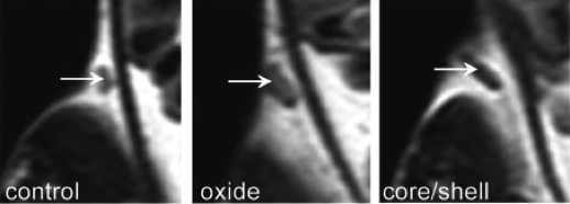
Figure 1: MRI images from iron-iron oxide core-shell nanoparticles injected into a mouse to enhance the contrast of a tumour.2
Using the leading edge of solution phase synthetic techniques, precise control over the nanoparticles and their magnetic properties can be achieved (Figure 2). In this project, well-defined nanoparticles with controlled crystalline domains will be studied for MPI. You will use transmission electron microscopy at one of the top microscopy facilities in Australia and be supervised by the director of the electron microscope unit, Professor Tilley. You will collaborate with leading researchers in MPI from Australia and internationally and work closely with a group of experts in nanoparticle synthesis. Overall, this work will tune nanoparticle size with precise synthetic control to optimise the magnetic properties of iron and iron oxide nanoparticles for MPI applications.
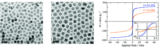
Figure 2: Transmission electron microscopy images of iron nanocubes and their magnetic properties for use in MPI.3
References
- Gloag, L., Mehdipour, M., Chen, D., Tilley, R. D. & Gooding, J. J. Advances in the Application of Magnetic Nanoparticles for Sensing. Adv. Mater. 31, 1904385 (2019).
- Cheong, S. et al. Simple synthesis and functionalization of iron nanoparticles for magnetic resonance imaging. Angew. Chem. - Int. Ed. 50, 4206–4209 (2011).
- Gloag, L. et al. Zero valent iron core–iron oxide shell nanoparticles as small magnetic particle imaging tracers. Chem. Commun. 56, 3504–3507 (2020).