
-
Lead contact: justin.gooding@unsw.edu.au
An unmet need in the field of biosensing is to develop technologies that can selectively detect target species at ultralow concentration levels.[1] This is important for disease diagnostics at earlier stages, infection detection and assessment of treatment efficacy. However, existing commercial technologies seldomly reach to clinically relevant concentrations, that is, sub-picomolar or lower. To develop ultralow detection limit biosensors, the three main challenges are having more sensitive transducers, mass transport of analyte molecules to the sensor in a reasonable timeframe and achieving good selectivity.[1-3]
In our research group, we have developed a suite of technologies that are amenable to commercialisation that can detect analyte species at femtomolar and lower levels. The three innovative technologies are (1) ultrasensitive electrochemical sensors with attomolar detection limits based on “dispersible electrodes” concept, (2) nanopore blockade sensors for detecting rare single molecules, and (3) single plasmonic nanoparticles based biosensors for the detection of many single molecules. These three technologies use the same strategy of making magnetic nanoparticles to collect the biomarker of interest rather than the conventional approach of making the biomarker to find the sensing surface. In addition to this, a common thread in these technologies is to use nanoparticles to confine the measurement volume to nanolitre or smaller such that a single molecule in that volume is an appreciable concentration. Ultimately, we aim to have these new sensors and tools operate in complex biological fluids for ultrasensitive bioanalysis applications.
![Figure 1. A major opportunity in biomedical sensors is to develop ultrasensitive biosensors that can selectively detect analyte of interest at ultralow concentration levels.[1]](/content/unsw-sites/au/en/science/our-schools/chemistry/our-research/our-research-groups/smart-materials-and-surfaces-research-group/sms-research/_jcr_content/root/responsivegrid-layout-fixed-width/responsivegrid-full-top/column_layout_1357290636/par_2_1_75/column_layout/par_1/accordion/accordion-item-0/column_layout/par_2_1_50/image_copy.coreimg.jpeg/1633920153383/2021-10-figure-1-0.jpeg) Figure 1. A major opportunity in biomedical sensors is to develop ultrasensitive biosensors that can selectively detect analyte of interest at ultralow concentration levels.[1] Smart Materials and Surfaces Group
Figure 1. A major opportunity in biomedical sensors is to develop ultrasensitive biosensors that can selectively detect analyte of interest at ultralow concentration levels.[1] Smart Materials and Surfaces GroupRelevant publications:
[1] Y.F. Wu, R.D. Tilley, J.J. Gooding. The Challenges and Solutions in Developing Ultrasensitive Biosensors, J. Am. Chem. Soc., 2019, 141, 1162-1170.
[2] J.J. Gooding, K. Gaus. Single molecule sensors: challenges and opportunities for quantitative analysis, Angew. Chem. Int. Ed., 2016, 55, 11354-11366.
[3] Y.F. Wu, D.T. Bennett, R.D. Tilley, J.J. Gooding. How Nanoparticles Transform Single Molecule Measurements into Quantitative Sensors. Adv. Mater., 2020, 32, 1904339.
1. Ultrasensitive electrochemical sensors based on “dispersible electrodes” concept.
The dispersible electrodes are gold-coated magnetic nanoparticles (Au@MNPs) that are modified with biorecognition molecules to facilitate the selective capture of a species of interest from sample matrix and rapid collection by a magnet at a macroscale electrode where quantification of captured analytes can be realised electrochemically.
This strategy has been successfully demonstrated by the detection of miRNA-21, selected as a cancer marker, in whole blood with 10 aM detection limits.[4] Our research indicated that the response time can be reduced significantly, and the detection limits can be lowered by more than 2500-fold, compared to that by a planar surface based sensor.
We are now advancing this technology through endeavours to have more robust nanoparticles, reliable electrochemical measurement techniques, and extended detection realm of this technology to other analytes such as DNA, proteins and other small molecules.[5-10].
![Figure 2. Schematic illustrating the steps involved for the detection of miRNA in blood enabled by nucleic acid hybridisation on gold-coated magnetic nanoparticles.[4]](/content/unsw-sites/au/en/science/our-schools/chemistry/our-research/our-research-groups/smart-materials-and-surfaces-research-group/sms-research/_jcr_content/root/responsivegrid-layout-fixed-width/responsivegrid-full-top/column_layout_1357290636/par_2_1_75/column_layout/par_1/accordion/accordion-item-0/column_layout_copy_c/par_2_1_50/image_copy.coreimg.jpeg/1633920749184/2021-10-figure-2-0.jpeg) Figure 2. Schematic illustrating the steps involved for the detection of miRNA in blood enabled by nucleic acid hybridisation on gold-coated magnetic nanoparticles.[4] School materials and surfaces group
Figure 2. Schematic illustrating the steps involved for the detection of miRNA in blood enabled by nucleic acid hybridisation on gold-coated magnetic nanoparticles.[4] School materials and surfaces groupRelevant publications:
[4] R. Tavallaie, J. McCarroll, M. Le Grand, N. Ariotti, W. Schuhmann, E. Bakker, R.D. Tilley, D.B. Hibbert, M. Kavallaris, J.J. Gooding. Nucleic acid hybridization on an electrically reconfigurable network of gold-coated magnetic nanoparticles enables ultrasensitive microRNA detection in blood, Nat. Nanotechnol., 2018, 13, 1066-1071.
[5] I.Y. Goon, L.M.H. Lai, M. Lim, R. Amal, J.J. Gooding. ‘Dispersible electrodes’: a solution to slow response times of sensitive sensors. Chem. Commun., 2010, 46, 8821-8823.
[6] K. Chuah, L.M.H. Lai, I.Y. Goon, S.G. Parker, R. Amal, J.J. Gooding. Ultrasensitive electrochemical detection of prostate-specific antigen (PSA) using gold-coated magnetic nanoparticles as ‘dispersible electrodes’. Chem. Commun., 2012, 48, 3503-3505.
[7] L.M.H. Lai, I.Y. Goon, K. Chuah, M. Lim, F. Braet, R. Amal, J.J. Gooding. The biochemiresistor: an ultrasensitive biosensor for small organic molecules. Angew. Chem. Int. Ed., 2012, 51, 6456-6459.
[8] D.F. Chen, Y.F Wu, S. Hoque, R.D. Tilley, J.J. Gooding. Rapid and ultrasensitive electrochemical detection of circulating tumor DNA by hybridization on the network of gold-coated magnetic nanoparticles. Chem. Sci., 2021, 12, 5196-5201.
[9] P. Moazzam, M. Myekhlai, A. Alinezhad, F.A. Alshawawreh, P. Bakthavathsalam, V.R. Gonçales, R.D. Tilley, J.J. Gooding. Ultrasensitive detection of programmed death-ligand 1 (PD-L1) in whole blood using dispersible electrodes. Chem. Commun., 2021, 57, 2559-2562.
[10] M. Mehdipour, L. Gloag, D.T. Bennett, S. Hoque, R. Pardehkhorram, P. Bakthavathsalam, V.R. Gonçales, R.D. Tilley, J.J. Gooding. Synthesis of gold-coated magnetic conglomerate nanoparticles with a fast magnetic response for bio-sensing. J. Mater. Chem. C, 2021, 9, 1034-1043.
2. Nanopore blockade sensors for detecting rare single molecules.
In this project, we integrate magnetic nanoparticles (MNPs) with an array of solid-state nanopores and develop a unique nanopore blockade sensor for the detection of single molecules. We can modify MNPs with analyte specific receptors and then apply them to capture specific analytes (e.g., protein markers) and block the nanopores but not translocate through the pore. Only when the MNP has captured a target protein molecule can the MNP remain blocking the nanopore under a reversal of magnet filed. As a result, stepwise current change will be observed, which enables the direct counting of the number of detected proteins for quantitative analysis. We have demonstrated that the nanopore blockade sensor can detect prostate-specific antigen (PSA), a prostate cancer relevant protein, at attomolar levels in blood.[11-13] For this project, the ongoing research is to investigate the potential of affinity-based differentiation of biomolecules under a magnetic field, increase the throughput of the technology for detecting many single molecules, push the detection limits to even lower concentrations, and combine with optical detection methods to develop electro-optical readout applied in the sensing system.
![Figure 3. Schematic illustration about the construction of nanopore blockade sensors for the ultrasensitive detection of proteins.[11]](/content/unsw-sites/au/en/science/our-schools/chemistry/our-research/our-research-groups/smart-materials-and-surfaces-research-group/sms-research/_jcr_content/root/responsivegrid-layout-fixed-width/responsivegrid-full-top/column_layout_1357290636/par_2_1_75/column_layout/par_1/accordion/accordion-item-0/column_layout_1839276028/par_2_1_50/image_1960308422.coreimg.jpeg/1633921898417/2021-10-figure-3.jpeg) Figure 3. Schematic illustration about the construction of nanopore blockade sensors for the ultrasensitive detection of proteins.[11]
Figure 3. Schematic illustration about the construction of nanopore blockade sensors for the ultrasensitive detection of proteins.[11]![Figure 4. A representative ionic current trace of nanopore blockade sensing showing the tracking of multiple sequential blocking and unblocking events at nanopore chips with an array of 3×3 nanopores.[11]](/content/unsw-sites/au/en/science/our-schools/chemistry/our-research/our-research-groups/smart-materials-and-surfaces-research-group/sms-research/_jcr_content/root/responsivegrid-layout-fixed-width/responsivegrid-full-top/column_layout_1357290636/par_2_1_75/column_layout/par_1/accordion/accordion-item-0/column_layout_1839276028/par_2_2_50/image.coreimg.jpeg/1633921883268/2021-10-figure-4-0.jpeg) Figure 4. A representative ionic current trace of nanopore blockade sensing showing the tracking of multiple sequential blocking and unblocking events at nanopore chips with an array of 3×3 nanopores.[11]
Figure 4. A representative ionic current trace of nanopore blockade sensing showing the tracking of multiple sequential blocking and unblocking events at nanopore chips with an array of 3×3 nanopores.[11]Relevant publications:
[11] K. Chuah, Y.F. Wu, S.R.C. Vivekchand, K. Gaus, P.J. Reece, A.P. Micolich, J.J. Gooding. Nanopore blockade sensors for ultrasensitive detection of proteins in complex biological samples, Nat. Common., 2019, 10, 2109.
[12] Y.F. Wu, K. Chuah, J.J. Gooding. Evaluating the sensing performance of nanopore blockade sensors: a case study of prostate-specific antigen assay. Biosens. Bioelectron., 2020, 165, 112434.
[13] Y.F. Wu, Y. Yao, S. Cheong, R.D. Tilley, J.J. Gooding. Selectively detecting attomolar concentrations of proteins using gold lined nanopores in a nanopore blockade sensor. Chem. Sci., 2020, 11, 12570-12579.
3. Single plasmonic nanoparticles-based biosensors for the detection of many single molecules.
In this project, we use commercial camera and a dark-field microscopy to analyse the localized surface plasmon resonance (LSPR) of single plasmonic nanoparticles. When a biochemical sensing reaction is occurred on a nanoparticle, the optical signature of the nanoparticle is altered thereby leading to a change in the LSPR of the nanoparticle.
By setting up a threshold, we can differentiate the unreacted and reacted nanoparticles, pushing this approach for the detection of individual biomarkers on individual nanoparticles.
The dark-field optical microscopy is used to monitor the LSPR arising from individual nanoparticles. The wide field nature of the measurement allows for simultaneous characterisation of a huge number of nanoparticles on one image graph, turning individual nanoparticles into single nanoparticle assays for parallel single molecule detection.
We are looking into new approaches to construct digital biosensors with plasmonic nanoparticles. With the help of computer algorithms, we can perform the LSPR analysis over several thousand gold nanoparticles in less than 1 s.[14].
Hence, a significant reduction in analysis time and enhanced throughput of single particle spectral analysis can be achieved for molecular counting and quantitation. By building up a core-satellite binding construct, sensitive detection of interleukin-6 (IL-6), associated with inflammatory and auto-immune processes of many diseases, has been demonstrated.[14-15]
Meanwhile, we are applying this technology to the detection of viral RNA, exploiting a new concept of performing quantitative analysis by counting many single-molecule detection events. To further advance this technology, we will keep developing robust nanoparticles, smart signalling approaches to induce a signal change while limiting the influence of nonspecific adsorption, advanced algorithms for rapid data processing and bioanalysis with complex biological samples.[16]
![Figure 5. Sensitive detection of IL-6 using single nanoparticle assay. Schematic of the immunoassay in the presence and absence of IL-6 and the relevant LSPR shifts of gold nanoparticles for the control and testing experiments.[14]](/content/unsw-sites/au/en/science/our-schools/chemistry/our-research/our-research-groups/smart-materials-and-surfaces-research-group/sms-research/_jcr_content/root/responsivegrid-layout-fixed-width/responsivegrid-full-top/column_layout_1357290636/par_2_1_75/column_layout/par_1/accordion/accordion-item-0/column_layout_copy_c_252959497/par_2_1_50/image_copy.coreimg.jpeg/1633922206645/2021-10-figure-5.jpeg) Figure 5. Sensitive detection of IL-6 using single nanoparticle assay. Schematic of the immunoassay in the presence and absence of IL-6 and the relevant LSPR shifts of gold nanoparticles for the control and testing experiments.[14]
Figure 5. Sensitive detection of IL-6 using single nanoparticle assay. Schematic of the immunoassay in the presence and absence of IL-6 and the relevant LSPR shifts of gold nanoparticles for the control and testing experiments.[14]Relevant publications:
[14] M. Sriram, B.M. Pouryousefi, P.R. Nicovich, D.T. Bennett, P.J. Reece, D.B. Hibbert, R.D. Tilley, K. Gaus, S.R.C. Vivekchand, J.J. Gooding. A rapid readout for many single plasmonic nanoparticles using dark-field microscopy and digital color analysis, Biosens. Bioelectron., 2018, 117, 530-536.
[15] B.P. Markhali, M. Sriram, D.T. Bennett, P.S. Khiabani, S. Hoque, R.D. Tilley, P. Bakthavathsalam, J.J. Gooding. Single particle detection of individual protein molecules using dark-field microscopy to avoid signals from nonspecific adsorption. Biosens. Bioelectron., 2020, 169, 112612.
[16] M. Sriram, K. Zong, S.R.C. Vivekchand, J.J. Gooding. Single nanoparticle plasmonic sensors. Sensors, 2015, 15, 25774-25792.
-
Lead contact: r.tilley@unsw.edu.au
Metal nanoparticles for active and stable catalysts in renewable energy storage.
There are several catalysis projects currently undertaken in the SMS research group:
1. Controlling nanoparticles structure for improved stability in oxygen evolution electrocatalysis.
The oxygen evolution reaction (OER) is crucial for the storage and conversion of H2 fuel and requires highly active and highly stable catalysts to drive it. Our expertise in nanoparticle synthesis has allowed us to create the most active and stable nanocatalysts for OER reported to date.1 We achieved this by synthesizing 3D branched Ru nanoparticles with structural features that both prevent dissolution and improve oxidation catalysis (Figure 1).2
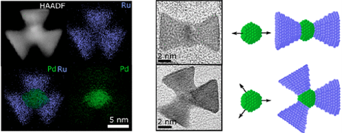 Figure 1: Energy dispersive X-ray spectroscopy elemental mapping of Pd-Ru branched nanoparticles and TEM images of individual nanoparticles. Models show the controlled direction of growth of Ru from Pd seed.3
In this project, Ru nanoparticles will be synthesized with low index facets which are critical for achieving stable reaction kinetics that prevent dissolution of Ru and enhance the catalytic activity.
This work will combine the development of synthetic methods to control the size, shape and composition of Ru-based nanocatalysts, with advanced characterisation using high-resolution transmission electron microscope (HRTEM) and also evaluation of their electrocatalytic performance.
This allows for the relationships between nanoparticle structure and catalytic performance to be fundamentally understood and tuned to create leading nanocatalyst materials.
- Gloag, L. et al. Three-Dimensional Branched and Faceted Gold-Ruthenium Nanoparticles: Using Nanostructure to Improve Stability in Oxygen Evolution Electrocatalysis. Angew. Chemie Int. Ed. 57, 10241–10245 (2018).
- Poerwoprajitno, A. R. et al. Formation of Branched Ruthenium Nanoparticles for Improved Electrocatalysis of Oxygen Evolution Reaction. Small 15, 1804577 (2019).
- Gloag, L. et al. A cubic-core hexagonal-branch mechanism to synthesize bi-metallic branched and faceted Pd-Ru nanoparticles for oxygen evolution reaction electrocatalysis. J. Am. Chem. Soc. 140, 12760–12764 (2018).
2. Synthesising strained Pt on metal nanoparticles for enhanced electrocatalytic activity in hydrogen fuel cells
Pt is the most catalytically active metal for fuel cell reactions, but it is also expensive. In order to convert to sustainable energy cells in a hydrogen economy, nanocatalysts need to be high-performing and use minimal amounts of scarce Pt. Strained Pt on the surface of a metal nanoparticle is a promising nanoparticle structure for highly active hydrogen evolution (HER) and oxygen reduction (ORR) electrocatalysis. As world-leaders in nanoparticle synthesis, we have been the first research group to create these via by a “bottom-up” approach involving the direct growth of Pt onto nanoparticles.1-3 Depositing Pt directly onto Ni nanoparticles creates highly strained Pt that maximises the specific and simultaneously minimising the amount of expensive Pt that is used to provide the highest mass activities reported to date (Figure 1).
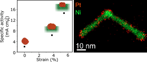 Figure 1: Relationship between strain and HER activity and elemental map of a Pt on Ni nanoparticle.2
In this project, nanoparticles will be decorated with small clusters of Pt atoms for use as high performance HER and ORR catalysts. The state-of-the-art electron microscopes provided by Electron Microscopy Unit, UNSW, will allow characterisation of complex nanocatalyst materials made of multiple metals with atomic-level precision. By controlling the position of Pt atoms on different metal nanoparticle structures, both electrocatalytic activity and stability will be optimised to create the most advanced and effective nanoparticle catalysts.
- Alinezhad, A. et al. Controlling Pt Crystal Defects on the Surface of Ni-Pt Core-Shell Nanoparticles for Electrocatalysts for Oxygen Reduction. ACS Appl. Nano Mater. 3, 5995-6000 (2020).
- Alinezhad, A. et al. Direct Growth of Highly Strained Pt Islands on Branched Ni Nanoparticles for Improved Hydrogen Evolution Reaction Activity. J. Am. Chem. Soc. 141, 16202–16207 (2019)
- Alinezhad, A. et al. Controlling hydrogen evolution reaction activity on Ni core–Pt island nanoparticles by tuning the size of the Pt islands. Chem. Commun. 57, 2788-2791 (2021)
3. In situ Transmission Electron Microscopy for nanoparticle catalyst design
The state-of-the-art electron microscopes managed by Professor Richard Tilley allow analysis of 3D structure, atomic arrangement and elemental composition of nanoparticles with unprecedented resolution at the nanoscale (Figure 1).1,2 As the first group in Australia to have in situ Transmission Electron Microscopy (TEM) facilities, we are paving the way for research into nanocatalyst design at the atomic level.
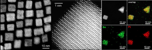 Figure 1: High angle annular dark field – scanning transmission electron microscopy images of nanocatalysts and energy dispersive x-ray spectroscopy mapping of elements showing spatial information of their structures and compositions.1
The structure of nanoparticles during catalysis is one of the hottest topics in fuel cell research at the moment. By understanding where and how gas molecules interact with nanocatalysts, the most active and stable catalyst materials can be designed from a fundamental basis. In this project, you will image real time changes to nanoparticle structure in the presence of different gases, to visually track interactions of molecules with catalytically active sites. The tomography capabilities of our microscopes allows for 3D visualisation of complex and intricate nanocatalyst structures that are leading particles for electrocatalytic reactions (Movie)
Figure 2: Tomographic reconstruction of a Au-Ni branched nanoparticle used for biomass oxidation.2
- Chen, H.-S. et al. Preserving the Exposed Facets of Pt3Sn Intermetallic Nanocubes During an Order to Disorder Transition Allows the Elucidation of the Effect of the Degree of Alloy Ordering on Electrocatalysis. J. Am. Chem. Soc. 142, 3231–3239 (2020).
- Chen, H.-S. et al. Role of the Secondary Metal in Ordered and Disordered Pt–M Intermetallic Nanoparticles: An Example of Pt3Sn Nanocubes for the Electrocatalytic Methanol Oxidation. ACS Catal. 11, 2235-2243 (2021).
- Poerwoprajitno, A. R. et al. Faceted Branched Nickel Nanoparticles with Tunable Branch Length for High Activity Biomass Oxidation Electrocatalysis. Angew. Chem. Int. Ed. 123, 15615-15620(2020).
-
Lead contact: justin.gooding@unsw.edu.au
Cell models are a fundamental element in all aspects of the literature surrounding cell biology. There is a shift in literature in modern literature whereby traditional 2D models are being replaced in favour of 3D models as these are more representative of the native extracellular matrix. 3D-bioprinting is an exciting technology that has the potential to produce 3D cell models in a reproducible and high throughput manner. We have developed a new 3D-bioprinter called the RASTRUM that is capable of high-throughput printing by utilising drop-on-demand technology to accurately create 3D cell models. The printer is comprised of a multivalve printhead with 2-axes of motion that prints a bioink and an activator that triggers the gelation to form a biomimetic matrix.
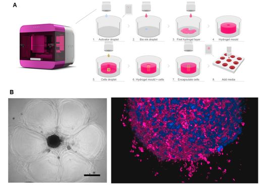 Figure 1: 3D bioprinting of spheroids using the RASTRUM 3D-bioprinter. (A) A schematic of the drop-on-demand system printing the cup. (B) Microscopy images of the printed spheroids in the cup.
In our project, we are developing a high-throughput drug screening method by using the RASTRUM to print a tissue like matrix to form a cup composed of an alginate-based bioink and calcium chloride activator (Fig. 1A). The printer deposits cells into the cup using optimised pressure and fluidic sheer to ensure optimal cell viability (Fig. 1B). This allows us to embed spheroids in 3D matrices with precise control over cell number and spheroid size.
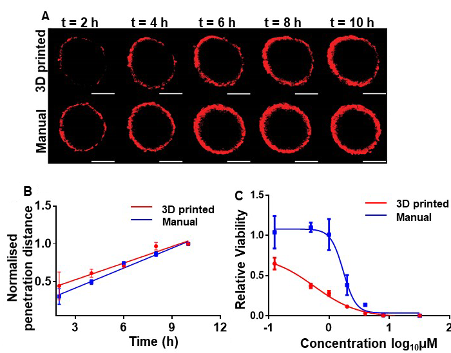 Figure 2. High-throughput screening data from the printed spheroids for (A) spheroid penetration and (B) spheroid viability.
We have demonstrated that this technology is suitable for high-throughput drug screening by printing a 96-well plate of spheroids and exposing them to different concentrations of doxorubicin for 72 h. The spheroids printed were of good quality and had drug penetration properties similar to that of manually produced spheroids (Fig. 2A and 2B), thereby proving that the printer-based assay was robust and met current standards. We also showed that the presence of a matrix greatly increased both the doxorubicin sensitivity of the spheroid (Fig. 2C) which highlights the differences between 2D and 3D cultures. This system has the capability to give end-users a reliable tool looking to design new assays as well as end-users in the clinical field looking for high-throughput screening.
With further development in regulating the drop-on-demand technology, we will be able to develop even more complex biological models by incorporating molecules that attract cells to certain locations within the hydrogel structure. This will allow us to explore the movement of cells to a chemical stimulus in 3D in a systematic manner with greater control over the microenvironment surrounding the cells. Furthermore, we will incorporate nanoscale sensors to measure the levels of extracellular biological molecules that indicate abnormal processes. This will evolve the printer to not only encompass high-throughput drug screening, but it will also become a powerful tool in disease prognosis and cancer relapse detection.
Relevant publications
1. Utama, R. H.; Atapattu, L.; O’Mahony, A. P.; Fife, C. M.; Baek, J.; Allard, T.; O’Mahony, K. J.; Ribeiro, J. C. C.; Gaus, K.; Kavallaris, M.; Gooding, J. J., A 3D bioprinter specifically designed for the high-throughput production of matrix-embedded multicellular spheroids. iScience 2020, 23, 101621.
2. Tan, V. T. G.; Nguyen, D. H. T.; Utama, R. H.; Kahram, M.; Ercole, F.; Quinn, J. F.; Whittaker, M. R.; Davis, T. P.; Justin Gooding, J., Modular photo-induced RAFT polymerised hydrogels via thiol–ene click chemistry for 3D cell culturing. Polymer Chemistry 2017, 8, 6123-33.
-
Lead contact: r.tilley@unsw.edu.au
Magnetic nanoparticles for cancer detection using Magnetic Particle Imaging
As the first to have a Magnetic Particle Imaging (MPI) instrument in Australia, at UNSW we are in a unique position to detect early stage tumours and cancerous cells with the most sensitive and precise imaging. The exceptional magnetic properties of iron and iron oxide nanoparticles make these ideal candidates for this state-of-the-art application. These key magnetic properties are dictated by the size, crystallinity and composition of the magnetic nanoparticles.1
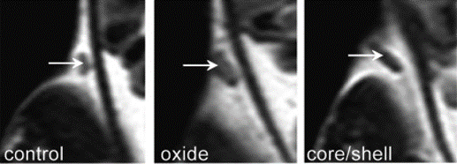 Figure 1: MRI images from iron-iron oxide core-shell nanoparticles injected into a mouse to enhance the contrast of a tumour.
Using the leading edge of solution phase synthetic techniques, precise control over the nanoparticles and their magnetic properties can be achieved (Figure 2). In this project, well-defined nanoparticles with controlled crystalline domains will be studied for MPI. You will use transmission electron microscopy at one of the top microscopy facilities in Australia and be supervised by the director of the electron microscope unit, Professor Tilley. You will collaborate with leading researchers in MPI from Australia and internationally and work closely with a group of experts in nanoparticle synthesis. Overall, this work will tune nanoparticle size with precise synthetic control to optimise the magnetic properties of iron and iron oxide nanoparticles for MPI applications.
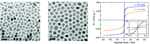 Figure 2: Transmission electron microscopy images of iron nanocubes and their magnetic properties for use in MPI.3
- Gloag, L., Mehdipour, M., Chen, D., Tilley, R. D. & Gooding, J. J. Advances in the Application of Magnetic Nanoparticles for Sensing. Adv. Mater. 31, 1904385 (2019).
- Cheong, S. et al. Simple synthesis and functionalization of iron nanoparticles for magnetic resonance imaging. Angew. Chem. - Int. Ed. 50, 4206–4209 (2011).
- Gloag, L. et al. Zero valent iron core–iron oxide shell nanoparticles as small magnetic particle imaging tracers. Chem. Commun. 56, 3504–3507 (2020).
-
Lead contact: justin.gooding@unsw.edu.au
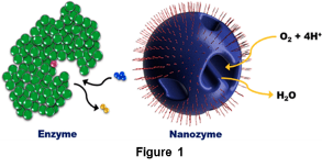
Enzymes have exceptional catalytic performance, which is largely related to their distinct structural features, such as having the active sites down a substrate channel to spatially separate the redox centre from the bulk solution environment. This provides control over the electronic properties, the chemical environment where the reaction proceeds and the transport of reactants/intermediates/products, including shuttling of intermediates to a second active site for cascade reactions.
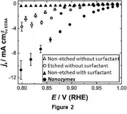
Inspired by enzymes, we have developed nanozymes: nanoparticles that mimic the tridimensional architecture of enzymes for electrocatalysis in a nanoconfined environment. We have made Pt-Ni nanoparticles that gave isolated channels when the Ni rich domains were etched away. The nanozymes outside surface remained passivated with a hydrophobic surfactant, allowing the electrocatalytic reaction to happen only inside the isolated nanochannels (Figure 1)1.
These nanozymes presented specific activity for the oxygen reduction reaction 3x higher than when the reaction happened at the surface outside the nanochannels (Figure 2). The higher activity is attributed to a different solution environment inside the nanochannels, with higher local concentration of H+compared to the bulk as shown by further changing the nanochannels dimensions and physicochemical modelling.2
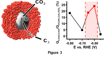
We also extended the concept of nanozymes to enable cascade reactions by having two different active sites located in close proximity in a nanoconfined environment. CO2 reduction reaction was chosen as the model reaction since it can be divided into two distinct processes: (1) CO2 reduction to CO and (2) CO reduction to higher order organic molecules. The nanozyme comprises a silver core surrounded by a porous copper shell. The first process (CO2 to CO) happens at the silver sites in the core at low overpotential and with high efficiency. The CO intermediate is then confined in the copper shell, being further reduced to higher organic molecules at the copper sites instead of diffusing away to the bulk solution3 Figure 3). The higher local concentration of CO at the confined copper sites promoted C-C coupling reactions, resulting in the formation of C3 products such as propanol at the lowest overpotentials reported to date. The rate of CO production can be modulated by switching the Ag with Pd at the core, resulting in higher structural stability and distinct products obtained at the copper sites.4
These results show how nanoconfinement has a profound effect on electrocatalysis. This concept can be further explored for different electrochemical reactions and by controlling the chemical environment inside the nanochannels to provide improved reaction kinetics and selectivity in the same way enzymes do.
1 T.M. Benedetti, C. Andronescu, S. Cheong, P. Wilde, J. Wordsworth, M. Kientz, R.D. Tilley, W. Schuhmann, J.J. Gooding, “Electrocatalytic nanoparticles that mimic the three-dimensional geometric architecture of enzymes: Nanozymes”, Journal of the American Chemical Society 140 (2018) 13449
2 J. Wordsworth, T. M. Benedetti, A. Alinezhad, R. D. Tilley, M. A. Edwards, W. Schuhmann, J. J. Gooding, “The importance of nanoscale confinement on electrocatalytic performance” Chemical Science 11 (2020) 1233
3 P. O'Mara, P. Wilde, T. M. Benedetti, C. Andronescu, S. Cheong, J. J. Gooding, R. D. Tilley, W. Schuhmann, “Cascade Reactions in Nanozymes: Spatially Separated Active Sites inside Ag Core-Porous Cu Shell Nanoparticles for Multistep Carbon Dioxide Reduction to Higher Organic Molecules” Journal of the American Chemical Society 141 (2019) 14093
4 P. Wilde, P.B. O'Mara, J.R.C. Junqueira, T. Tarnev, T.M. Benedetti, C. Andronescu, Y-T. Chen, R. D. Tilley, W. Schuhmann, J. J. Gooding, “Is Cu instability during the CO2 reduction reaction governed by the applied potential or the local CO concentration?” Chemical Science 12 (2021) 4028-4033
-
Lead contact: justin.gooding@unsw.edu.au
Wearable sensors have received increasing attention in medicine, safety, and military. Such devices can monitor vital chemical parameters of individuals continuously or record exposures to a situation over time. This capability allows a fast response to a change in health or safety condition. For instance, our research group has previously demonstrated a wearable paper-based sensor that changes colour according to sun exposure. The sensor works by employing a polyvinylpyrrolidone polymer as a binder for titanium oxide particles and food dyes. The titanium oxide works as a photocatalyst that degrades the dyes with exposure to UV radiation. The discolouration can be observed by the naked eye, informing the user about overexposure to UV radiations.
![Change in colour versus time of exposure to UV radiation. [2]](/content/unsw-sites/au/en/science/our-schools/chemistry/our-research/our-research-groups/smart-materials-and-surfaces-research-group/sms-research/_jcr_content/root/responsivegrid-layout-fixed-width/responsivegrid-full-top/column_layout_1357290636/par_2_1_75/column_layout/par_1/accordion/accordion-item-3/column_layout_copy_6/par_2_1_50/image_copy.coreimg.png/1633926029701/2021-10-index.png) Change in colour versus time of exposure to UV radiation. [2]
Change in colour versus time of exposure to UV radiation. [2]Following the current trend of continuous fitness monitoring systems, wearable sensors technology are equally compatible with mobile smartphones. In this context, the ability to track progress and share data is a central pivot to the democratisation of health care monitoring. The goal is to confer individuals with the capability of observing and managing the evolution of critical parameters that can affect their wellbeing.
The pursuit of wearable sensors started in hospitals. The motivation was to diminish the long time needed for receiving data of samples collected from a patient and sent to a centralised laboratory for analysis. In critical care situations, this workflow is not compatible with the tight time frames required to make critical therapeutic decisions. Sensor researchers were pivotal to this breakthrough. The development of the field continued with hand-held devices that patients can now use at home to reduce medical visits. Glucose monitors, in particular, have been a phenomenal success story in this regard. Nowadays, with the need for health-dedicated wearable sensors for a range of analytes other than glucose, there are important questions that our research group wants to address. Such questions involve new science that needs to be understood and characterised to allow a long and continuous use of wearable sensors. In particular, there is a strong need for new approaches that can improve our capability to prevent nonspecific adsorption of molecules on the biorecognition interface and for the development of calibration-free sensors. Addressing both questions may allow the development of long-term wearable devices where no intervention from the user is required. This is a field receiving increasing attention of academics and companies.
![Figure – Interfaces that can improve our capability of avoiding nonspecific adsorption are vital for long-term operation of wearable devices [4].](/content/unsw-sites/au/en/science/our-schools/chemistry/our-research/our-research-groups/smart-materials-and-surfaces-research-group/sms-research/_jcr_content/root/responsivegrid-layout-fixed-width/responsivegrid-full-top/column_layout_1357290636/par_2_1_75/column_layout/par_1/accordion/accordion-item-3/column_layout_copy_6_1974929931/par_2_1_50/image.coreimg.png/1633926072915/2021-10-index2.png) Figure – Interfaces that can improve our capability of avoiding nonspecific adsorption are vital for long-term operation of wearable devices [4].
Figure – Interfaces that can improve our capability of avoiding nonspecific adsorption are vital for long-term operation of wearable devices [4].[1] E. Bakker, J.J. Gooding. Wearable Sensors – An Exciting Area of Research for Sensors Scientists. ACS Sensors. 2016, 1, 834-834.
[2] P.S. Khiabani, A.H. Soeriyadi, P.J. Reece, J.J. Gooding. Paper-Based Sensor for Monitoring Sun Exposure. ACS Sens. 2016, 1, 775−780.
[3] J.J. Gooding. Finally, a simple solution to biofouling. Nature Nanotechnology 2019, 14, 1089–1090.
[4] C. Jiang, M.T. Alam, S.M. Silva, S. Taufik, S. Fan and J.J. Gooding. Unique Sensing Interface That Allows the Development of an Electrochemical Immunosensor for the Detection of Tumor Necrosis Factor α in Whole Blood. ACS Sens. 2016, 1, 1432.