
SEM - TFS Verios 5 XHR
High Resolution SEM imaging, windowless light element EDS analysis and large area EDS analysis

Description
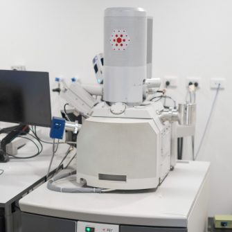
The Thermofisher Verios XHR is a field emission scanning electron microscope capable of achieving sub-nanometre resolution. It has the capability to operate at a wide range of beam conditions, from 500V to 30kV, with beam current ranging from a few pA to many nA.
A wide range of electron detectors are available on the microscope, for both secondary and backscatter imaging. Adding to this, a large piezo stage allows accurate sample movement, enabling repeatable and reliable measurements. A cryo-stage can also be attached to the microscope, so it is possible to study samples under low temperature conditions.
The Verios is also equipped with 3 EDS detectors from Oxford Instruments: a large area conventional detector (Ultim MAX Infinity), a pole piece level detector (Unity BEX) and a specialised windowless detector optimised for low-energy X-ray detection (Ultim Extreme Infinity). This makes the Verios an extremely versatile instrument, with the capability to perform both ultra-high-resolution imaging and chemical analysis on many different types of specimens. For air-sensitive samples, inert transfer from a compatible glove box or the CleanMill ion mill is possible using a dedicated CleanConnect sample transfer system.
Specifications
-
- 0.35kV to 30kV, 0.8pA to 100nA; landing energy can be as low as 20eV with beam deceleration.
-
- 0.8nm at 1kV, 0.7nm at 15kV and 0.6nm 30kV (STEM).
-
- IR chamber camera & Nav-CamTM for sample navigation
- Everhart-Thornley SE detector (ETD)
- In-lens detector (TLD)
- In-column BSE detectors (MD and ICD)
- Retractable concentric solid-state BSE detector (DBS)
- Retractable segmented STEM detector
-
- Hybrid high solid angle EDS-BSE detector (Oxford Unity BEX)
- Windowless EDS detector (Oxford Ultim Extreme Infinity)
- Large area conventional SDD-EDS detector (Oxford Ultim MAX Infinity 170mm2)
-
- CryoMAT low temperature stage
- CleanConnect inert sample transfer system
Applications
- Battery and Photovoltaic materials
- Nanoparticles
- Metals and alloys
- Semiconductors
- Composite materials
Capabilities
- High Resolution SEM imaging
- Large area chemical analysis
- Light element chemical analysis
- Low temperature SEM imaging
- STEM
- Large area SEM imaging (MAPS)
Publishing Microscopy Data Acquired on the Thermo Fisher Scientific Verios 5 XHR
-
Acknowledgement:
“The authors acknowledge the facilities and the scientific and technical assistance of Microscopy Australia at the Electron Microscope Unit (EMU) within the Mark Wainwright Analytical Centre (MWAC) at UNSW Sydney.”
Credit EMU staff: Feel free to mention EMU staff who have assisted you with your work! If staff have been involved with your work beyond basic training and support (e.g., project design, complex data/image processing, independent imaging/analysis, manuscript preparation), it may be appropriate to discuss co-authorship with the relevant staff and your supervisor.
Don’t forget to email the EMU lab manager with a copy of your publication to claim 2 hours of free microscopy time.
Funding patners


Instrument location
Electron Microscope Unit
B85, Basement
June Griffith Building (F10)
UNSW Sydney, NSW 2052
Access – To get access to the SEM, either for training, collaboration or staff assisted work, please fill in the Verios Access Form
Associate Professor Shery Chang
Associate Director Electron Microscopy
-

Email
