Early and reliable detection of Parkinson's disease in cerebrospinal fluid
New detection method developed by Sydney scientists could enable early, accurate diagnosis for Parkinson’s disease.
Published on the 19 Feb 2021 by Sue Min Liu

New detection method developed by Sydney scientists could enable early, accurate diagnosis for Parkinson’s disease.

UNSW Sydney medical researchers create a new diagnostic method for Parkinson’s disease that combines a single-molecule counting technique with a rapid amplification assay to detect alpha-synuclein – a promising biomarker for the disease. This technique will allow clinicians to detect Parkinson’s disease at early stages before the onset of clinical signs, which will lead to better outcomes for the patients.
Clumping of the protein alpha-synuclein in cells of the nervous system is a hallmark of Parkinson’s disease, as well as other neurological disorders, including Lewy body dementia, and multiple system atrophy. With no specific biomarkers to reliably diagnose Parkinson’s disease, clinicians rely on a combination of signs and symptoms, including tremors and responsiveness to specific treatments. The only definite way to diagnose Parkinson’s disease, at present, is at autopsy.
As reported in a paper published in Angewandte Chemie International Edition online recently, a new diagnostic strategy enables the analysis of protein aggregates in cerebrospinal fluid to detect alpha-synuclein, and discriminate between Parkinson’s disease and other neurodegenerative diseases.
Parkinson’s disease is driven by highly organised strings of alpha-synuclein protein, called fibrils. The fibrils have the capacity to seed protein clumps in cells, and spread to other cells, ultimately interfering with normal functions of the brain. The research team show that, even at very low concentrations, these fibrils can be detected with a compact and simple to operate 3D-printed microscope developed previously by the study co-leads Dr Emma Sierecki and Dr Yann Gambin at UNSW Medicine and Health’s Single Molecule Science.
“Single-molecule counting allows precise quantitation and fingerprinting of protein aggregates, including size and reactivity of individual aggregates to identify unique signatures for each patient,” Dr Gambin said.
To enhance sensitivity and reliability of detection, the team coupled single-molecule detection with a short one-step assay to amplify alpha-synuclein fibrils. This method can detect fibrils in a little over five hours. Other molecular detection methods are under development by other research teams, but most of these require multiple steps to amplify alpha-synuclein requiring more than 50 hours.
“Alpha-synuclein aggregates can be quite different and do not react the same way to amplification. This method will make it easier to distinguish between Parkinson’s disease and other diseases associated with alpha-synuclein aggregates. The amplification step also ensures that we are actually detecting alpha-synuclein,” Dr Sierecki said.
The team are excited that the method works in complex and clinically relevant samples like cerebrospinal fluid, and with the funding they recently secured from the Michael J. Fox Foundation and the Shake It Up Australia Foundation, their plan is to test and adapt the assay for detection of alpha-synuclein fibrils in biofluids that can be collected with less invasive procedures, including blood, urine and saliva.
“Hopefully we are getting the quantitative tools that are currently missing in Parkinson’s disease diagnostics. This new method can also be used during clinical trials to monitor disease progression, which currently rely on improvement of symptoms, with little quantitative biochemical analysis,” said Dr Sierecki.
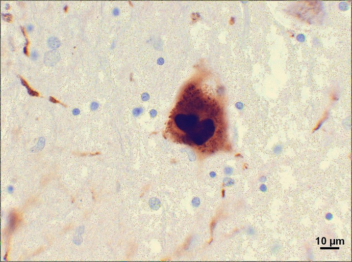
[Feature image: Micrograph of clumps of alpha-synuclein protien in the brain of a patient with Parkinson's disease by Suraj Rajan Wikimedia Commons, CC-BY-SA-3.0]
Contact Name: Sue Min Liu
Email: sue.liu@unsw.edu.au