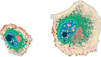
A team of researchers from Helmholtz Munich, the University of Zurich and UNSW Sydney have developed a powerful new tool for analyzing the spatial organization of cells using high-resolution fluorescence microscopy. In a paper published in Nature Methods, they describe the development of CAMPA (Conditional Autoencoder for Multiplexed Pixel Analysis), a deep-learning framework that allows the study of subcellular organization in both healthy cells and those engineered with specific mutations.
Prof Fabian Theis, corresponding author from Helmholtz Munich, explains their reasoning. “Foundation models such as large language models are the next big thing in machine learning and AI. A long-term goal of our research is to build a foundation model of the cell. CAMPA transfers recent work from us and others on AI for single cell gene expression variation towards learning about spatial subcellular structure, which constitutes another step towards such a model,” he says.
“Our goal was to develop an automated method for the quantitative analysis of subcellular organization, with the potential to facilitate analysis of high-throughput screens,” says study co-lead, Dr Hannah Spritzer from Helmholtz Munich.
Subcellular organization is crucial for cell function. To identify consistent subcellular landmarks present in every cell across a myriad developmental and experimental conditions, CAMPA uses a conditional variational autoencoder, which is an unsupervised deep learning method. These landmarks can be used to create interpretable cellular fingerprints, allowing researchers to identify similarities and differences between cells under different conditions, and enhancing our understanding of cellular processes. Using CAMPA, researchers can characterize different growth phases and functional disruptions in terms of their subcellular phenotypes, meaning that this can be a powerful tool for cellular phenotypic screening.
“With CAMPA, we can now consistently identify the different organelles and other structures in cells directly from highly multiplexed images, which greatly accelerates our ability to gain insight from these complex datasets,” highlights Dr Scott Berry, study co-first author from the University of Zurich and UNSW.
“This can be a game-changer for drug discovery. With CAMPA, we can now use our iterative indirect immunofluorescence imaging platform to compare, at detailed subcellular resolution, the effects of drugs, which can give unprecedented rich insights into their mechanism of action, and ultimately their therapeutic potential,” says Prof Lucas Pelkmans, corresponding author, also from the University of Zurich.
Ultimately, CAMPA can help to build a queryable atlas of intracellular variation which would be instrumental in uncovering the rules by which spatial context shapes genome activity across multiple scales.
Article adapted from Helmholtz Munich Newsroom: CAMPA: A Powerful Deep Learning Framework for Understanding Subcellular Organization
See also Research Briefing of this study by Hannah Spitzer and Fabian Theis in Nature Methods.