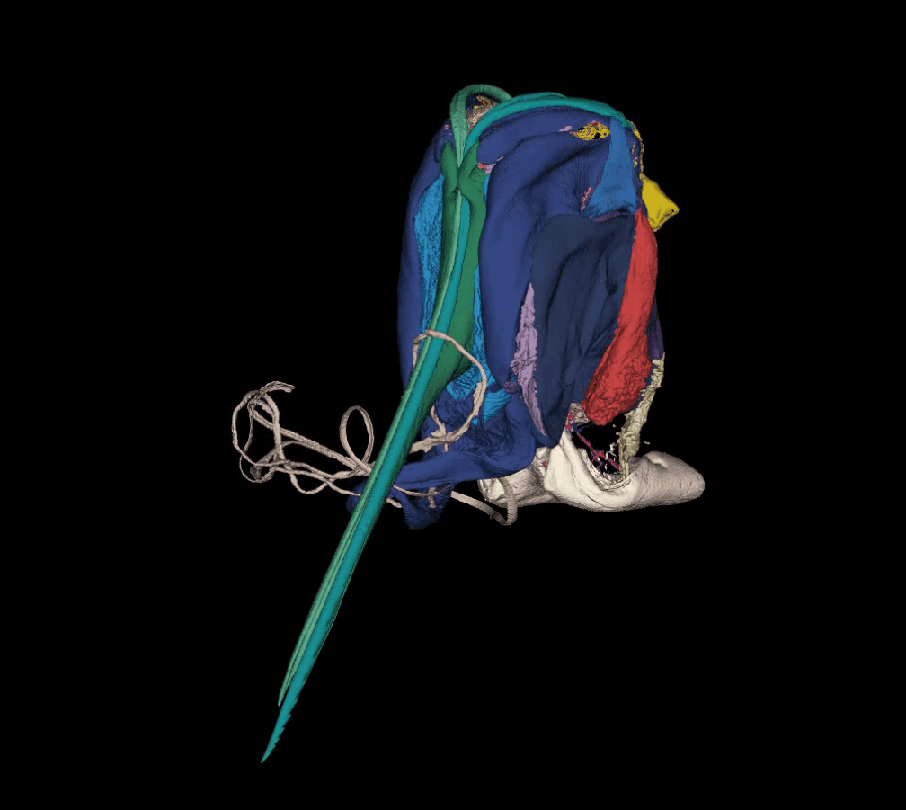
Groundbreaking new high-resolution imagery of bee stingers is paving the way for the next generation of “micro" medical devices.

The 3D scans and reconstructions produced by UNSW Canberra researchers will help develop prototypes for tiny devices that can stay firmly attached to the skin and allow for more targeted drug delivery.

Recently published in the iScience journal, the 3D reconstructions show the unique properties of a bee stinger, including numerous barbs which are thought to be a key part of why stingers get left behind after stinging. Once separated from the bee the stinger continues to function autonomously and works its way deeper into the skin whilst also pumping venom.

According to UNSW Canberra's lead researcher, Associate Professor Sridhar Ravi the autonomous delivery mechanism of the bee stinger has numerous unique features that can be used in the development of small-scale and minimally intrusive medical devices.

“We have never before produced images with this level of detail, and they have given us tremendous new insights into the functions of the bee stinger. These latest findings have laid the foundations for the development of new types of medical devices,” Associate Professor Ravi said.

“Because of these clearer and more precise images we have uncovered opportunities in the areas of medical micro drilling, micro pumps and much more targeted drug delivery.”

“There is also the possibility of developing improved “anchoring” methods that will allow medical devices or adhesive patches to hold on to the skin without the need for chemical adhesives which can cause irritation or be unviable on moist surfaces, like the inside of the body.

“Previous studies have shown that a bee stinger is very good at piercing skin with minimal force, but it is very hard to remove once it is embedded. This is a really useful property for medical devices that need to be very precisely inserted without damaging surrounding tissues.”

The 3D reconstructions have already led to the UNSW Canberra research team developing prototypes of devices that can simulate the unique piercing and pumping actions of the bee stinger.

The project's other primary researcher, Dr Fiorella Ramirez Esquivel said that because a bee stinger is so small, at approximately 2mm in length, the research team had to use a combination of techniques to observe the stinger and decode how it works.

“The 3D reconstructions have been fantastic because they allowed us to 3D print the whole stinger and blow it up to a scale where we can move all the parts around to figure out how they work together. High speed filming the stinger in action was also a significant challenge but it has been instrumental in understanding how it functions,” Dr Ramirez Esquivel said.

Dr Ramirez Esquivel said that the evolution of the bee's stinger is a great example of how humans must sometimes refer to other animal and plant species in order to make progress.

“Bee stingers are incredibly complex structures with numerous moving components that also happen to be incredibly effective and efficient at what they do,” Dr Ramirez Esquivel said.

“A bee's stinger must be able to firstly pierce skin without buckling and it must safely detach and coordinate the muscular contractions that generate stinging. This means both working itself deeper into tissue and pumping venom quickly and efficiently.

“The bee stinger is such a complex organ. The more we look into it the more we find amazing intricacies related to how it does its job. There are endless possibilities for bio-inspired design contained within this tiny little machine.

“As advanced manufacturing makes strides in what it is possible for us to make, natural materials like the insect cuticle will become more and more relevant to the design of soft robots and microdevices.”

Images from this research project can be downloaded from this link: https://photos.app.goo.gl/UGztRam1Rc8kU3ku7


Animated 3D scans of a bee's stinger have provided greater detail about how the unique organ works. Source: Dr Fiorella Ramirez Esquivel