Nikon Ti2 Biosciences

Description
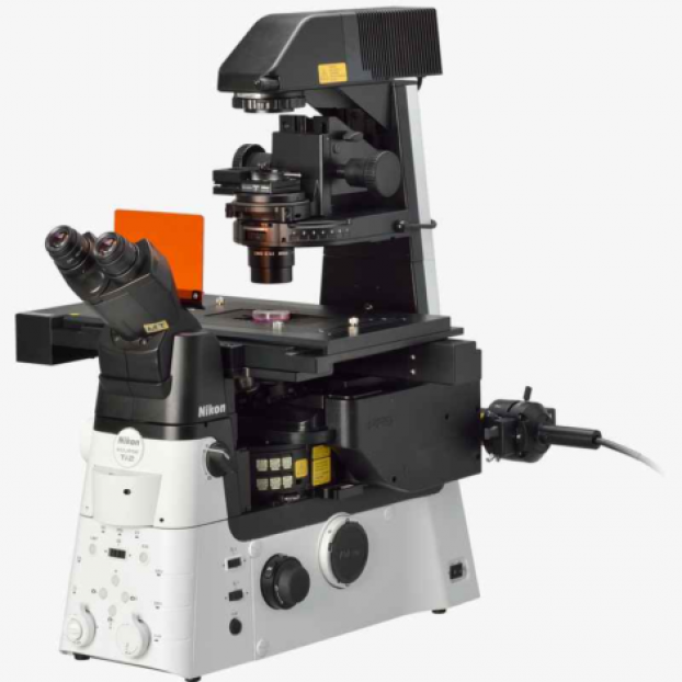
The Nikon Eclipse Ti2-E is an inverted widefield microscope known for its large field of view, high-quality optics, and advanced focus stabilization with Nikon's Perfect Focus System. It features Nikon JOBS software for automated experimental protocols and real-time analysis, along with a CoolLED light source for fast ratiometric imaging of calcium dynamics.
Specifications
-
Magn.
N.A.
Corrections
Immersion
W.D.
Misc.
4x
Plan Fluor
Air
17.2 mm
10x
0.3
Plan Fluor
Air
16 mm
Ph1
20x
0.5
Plan Fluor
Air
2.1 mm
Ph1
20x
0.75
Plan Apo VC
Air
1 mm
DIC N2
60x
1.2
Plan Apo VC
Oil
0.31 mm
DIC N2
20x
0.45
S Plan Fluor ELWD
air
8.2 mm
DIC N1
-
- LED DIA Illumination
- CoolLED with 340 nm, 380 nm and 430/60 nm visible range illumination
-
FURA
DAPI/FITC/
TRITC/CY5
(QUAD)
GFP-8
C-FL-C mCherry HQ
C-FL-C Cy5 HQ
Mirror
Peak 409
Peak 409/493/573/652
505
600
660
Excitation
Peak 378/474/554/635
460-500
550-590
590-650
Emission
510-560
608-683
663-738
-
- DAPI: 405-485 nm
- FITC: 515-555 nm
- TRITC: 590-650 nm
- Cy5: LP 670 nm
-
Hamamatsu Orca Flash 4.0 C11440 sCMOS Camera
- Low readout noise (Standard scan): 1.5 electrons rms, 0.9 electrons
- High speed readout (Rapid rolling, Full resolution): 40 frames/s
- Large field of view: 13.312 mm (H)×13.312 mm (V)
- High resolution: 2048 (H)×2048 (V)
- High quantum efficiency: 82 % (Peak QE)
Funding agency
- Luminesce Alliance (OHMR)
Applications
- Live-cell imaging
- Brightfield/DIC/Phase Contrast Microscopy
- Widefield Fluorescence Microscopy
- Calcium Imaging
- Automated Experiments via JOBS
Instrument location
Katharina Gaus Light Microscopy Facility
Biosciences Building (E26), Room 2013
UNSW Sydney, NSW 2033
Phone: 02 9065 5306
Email: KGlmf@unsw.edu.au
Dr Alex Macmillan
-
Email
alex.macmillan@unsw.edu.au
Dr Michael Carnell
-
Email
m.carnell@unsw.edu.au
Parent facility
Explore more instruments, facilities & services
Our infrastructure and expertise are accessible to UNSW students and staff, external researchers, government, and industry.


