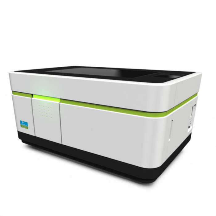
Perkin Elmer Operetta CLS

Description
The Operetta CLS is a high-content imaging system with a spinning disk confocal that supports rapid imaging of biological samples. Its automated capabilities enable high-throughput analysis, making it well-suited for applications such as phenotypic screening, live-cell imaging, and drug discovery research.
Specifications
-
- 5x magnification features a numerical aperture of 0.16, uses dry immersion, and offers a working distance of 12.1 mm, corrected for a 0.17 mm glass bottom.
- 20x magnification (1) has a numerical aperture of 0.4, with dry immersion and a working distance of 8.28 mm, corrected for glass bottoms ranging from 0 to 1.5 mm.
- 20x magnification (2) provides a numerical aperture of 0.8, utilises dry immersion, and has a working distance of 0.55 mm, corrected for a 0.17 mm glass bottom.
- 20x magnification (3) offers a numerical aperture of 1.0, uses water immersion, and has a working distance of 1.7 mm, corrected for a 0.17 mm glass bottom.
- 40x magnification comes with a numerical aperture of 1.1, utilises water immersion, and provides a working distance of 0.62 mm, corrected for a 0.17 mm glass bottom.
-
The system includes LED excitation at 365 nm, 405 nm, 440 nm, 475 nm, 510 nm, 550 nm, 630 nm, and 660 nm, as well as an LED for transmitted white light.
-
Filter Type
Wavelength Range
Excitation Filter
BP 355 nm – 385 nm
BP 390 nm – 420 nm
BP 435 nm – 460 nm
BP 460 nm – 490 nm
BP 490 nm – 515 nm
BP 500 nm – 550 nm
Emission Filter
BP 430 nm – 500 nm
BP 570 nm – 620 nm
BP 530 nm – 560 nm
BP 615 nm – 645 nm
BP 650 nm – 675 nm
BP 655 nm – 705 nm
BP 685 nm – 760 nm
BP 570 nm – 650 nm
-
The Hamamatsu Orca Flash 4.0 V2 camera features a CMOS image sensor for scientific measurement, with a maximum pixel format of 2048 x 2048, a pixel size of 6.5 µm (H) x 6.5 µm (V), a bit depth of 16 bits, and a sensitivity and speed of 100 fps at the 2048 x 2048 format.
Applications
- Phenotypic screening
- Live-cell imaging
- Drug discovery research
Instrument location
Katharina Gaus Light Microscopy Facility
Lowy Cancer Research Centre (C25),
Lower Ground, Room LG06,
UNSW Sydney, NSW 2033
Phone: 02 9065 5306
Email: KGlmf@unsw.edu.au
Dr Alex Macmillan
-
Email
alex.macmillan@unsw.edu.au
Dr Michael Carnell
-
Email
m.carnell@unsw.edu.au
Parent facility
Explore more instruments, facilities & services
Our infrastructure and expertise are accessible to UNSW students and staff, external researchers, government, and industry.


