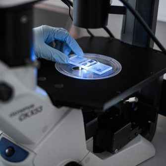
Leica DM IL LED Inverted Laboratory Microscope

Description
The Leica DM IL LED is an inverted laboratory microscope ideal for cell culture, micromanipulation, fluorescent immunostained specimens, and routine live cell examinations. It offers phase contrast, modulation contrast, and fluorescence imaging. The Leica EL6000 fluorescent light source enhances fluorescence imaging by connecting via a liquid light guide, keeping heat away from the specimen and microscope, and using a long-lasting mercury metal halide bulb. This microscope has two filters suitable for the common fluorophores DAPI and FITC. This microscope is also equipped with the Leica K3C is a high-performance monochrome microscope camera designed for a variety of applications in life sciences, industry, and clinical settings. It offers excellent dynamic range and high frame rates, making it ideal for imaging challenging samples with both brightfield and fluorescence microscopy.
Specifications
-
- Inverted laboratory microscope
-
- Brightfield LED illumination, 5 W
-
- Phase contrast, modulation contrast, fluorescence
-
Leica EL6000
- Leica EL6000 External light source for fluorescence excitation
- Liquid light guide connection
- Mercury metal halide bulb with over 2000 hours lifetime
- Integrated fast shutter (6 ms response time)
- Attenuator to reduce photo bleaching
-
Leica K3C
- Sensor Type: CMOS
- Resolution: 3072 x 2048 pixels
- Pixel Size: 2.4 μm x 2.4 μm
- Exposure Range: 1 ms - 10 s
- Frame Rate: 15 fps (software triggered)
- Bit Depth: 32-bit RGB, 12-bit mono
- Binning Options: 2x2
- Optical Filter: 650 nm IR cut-off filter
Publishing Microscopy Data Acquired on the Leica DM IL LED with Leica EL6000 and K3C Camera
-
-
- Live cell details (e.g. confluency, cell type, passage)
- Fixing methods
- Staining methods and fluorophores used
- Mounting on slides
-
- Manufacturer: Leica
- Model: DM IL LED
- Type: Brightfield Inverted Laboratory Microscope
- Fluorescence Light Source: Leica EL6000
- Camera: Leica K3C
-
- Illumination type (Brightfield LED or fluorescence)
- Contrasting methods used (phase contrast, modulation contrast, fluorescence)
Acknowledgement:
“The authors acknowledge the facilities and the scientific and technical assistance of Cell Culture Facility (CCF) within the Mark Wainwright Analytical Centre (MWAC) at UNSW Sydney.”
Credit MWAC CCF staff: Feel free to mention MWAC CCF staff who have assisted you with your work! If staff have been involved with your work beyond basic training and support (e.g., project design, complex data/image processing, independent imaging/analysis, manuscript preparation), it may be appropriate to discuss co-authorship with the relevant staff and your supervisor.
-
Applications
- Cell culture monitoring
- Micromanipulation
- Immunostained specimen documentation
- Routine live cell examinations
- Fluorescence imaging
Instrument location
MWAC Cell Culture Facility
Laboratory 524D Science and Engineering Building (SEB) – E8
Kensington, UNSW, Sydney,
NSW, 2052
Email: CCFAdmin@unsw.edu.au
Access
-
Email
CCFAdmin@unsw.edu.au
Parent facility
Explore more instruments, facilities & services
Our infrastructure and expertise are accessible to UNSW students and staff, external researchers, government, and industry.


