Olympus R1 Cell Counter

Description
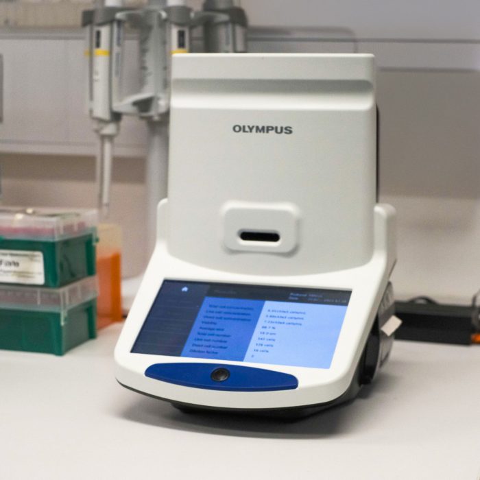
The Olympus R1 Cell Counter is designed to streamline and enhance the cell counting process. It provides fast, accurate counting results for a variety of cell types by automatically de-clustering clumped cells, performing size- and roundness-based sorting, and identifying live/dead cells. The R1 cell counter can confirm cell counts prior to experimental data acquisition, quality checks, passaging, or induction of differentiation. It also features a liquid lens autofocusing system for clear cell images and efficient cell culture data management.
Specifications
- Measurement Parameters: Confluency, cell count, and morphology.
- Output Information: Total/live/dead cell concentration, total/live/dead cell number, viability, average cell size.
- Supported Cell Types: Adherent cells, cell lines, spheroids, organoids.
- Sample Holders/Observable Range: Compatible with Olympus single use hemocytometers
- Imaging System: Automated cell culture measurements with intuitive interface.
- Cell Concentration Range: 5 × 10^4 – 1 × 10^7 cells/mL.
- Cell Diameter Range: 3–60 μm (recommended: 8–30 μm).
- Focusing Mode: Automatic/manual focusing with liquid lens.
- Adjustable Parameters: Size, roundness, dilution factor, noise reduction, delustering.
Publishing Microscopy Data Acquired on the Olympus R1 Cell Counter
-
-
- Cell name
- Cell passage number
- Culture vessel
- Viability solution dilution factor
- Culture type (suspension, adherent, spheroid)
-
- Manufacturer: Olympus
- Model: R1
- Type: Cell Counter
-
- Measurement parameters (confluency, cell count, morphology, viability)
-
- Adjustments to contrast/brightness
- Protocol parameters ( Size, roundness, dilution factor, noise reduction, delustering).
-
- Levels adjustments
Acknowledgement:
“The authors acknowledge the facilities and the scientific and technical assistance of Cell Culture Facility (CCF) within the Mark Wainwright Analytical Centre (MWAC) at UNSW Sydney.”
Credit MWAC CCF staff: Feel free to mention MWAC CCF staff who have assisted you with your work! If staff have been involved with your work beyond basic training and support (e.g., project design, complex data/image processing, independent imaging/analysis, manuscript preparation), it may be appropriate to discuss co-authorship with the relevant staff and your supervisor.
-
Applications
- Life Sciences
- Biomaterials
- Medical Sciences
- Cell Culture Research
- Drug Discovery
- Bioprocess Applications
Instrument location
MWAC Cell Culture Facility
Laboratory 524D Science and Engineering Building (SEB) – E8
Kensington, UNSW, Sydney,
NSW, 2052
Email: CCFAdmin@unsw.edu.au
Access
-
Email
CCFAdmin@unsw.edu.au
Parent facility
Explore more instruments, facilities & services
Our infrastructure and expertise are accessible to UNSW students and staff, external researchers, government, and industry.


