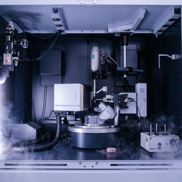
Bruker D8 Venture

Description
The Bruker D8 Venture X-ray diffractometer is a state-of-the-art instrument suitable for protein crystal diffraction. It is fitted with the following features: Photon III detector, Automatic ‘2 click’ AGH Kappa goniometer, Diamond IµS copper source, Oxford Cryo-stream, optional use of the ISX plate goniometer.
Specifications
-
Bruker Photon III 28 mixed mode x-ray detector
This detector can work in both photon counting and integration modes allowing high signal-to-noise ratios and accurate intensities. There is no detector noise, enabling high linearity and excellent quality data. Typical 'still' frames are in the order of 10 second exposures, with effectively instantaneous read-out. -
Automatic Goniometer Heade (AGH) kappa goniometer
The AGH enables 'two click' optical centering of the sample in a semi-automatic mode similar to Synchrotron MX beamlines. The goniometer is fitted with fast, high precision piezoelectric motors and has a built-in magnetic mount that accepts standard mounting pins. The Bruker software allows full control of the kappa functions of the goniometer, and the AGH works with low temperature (100 K) operation. -
Diamond IµS copper
The UNSW Bruker diffractometer has a Diamond IµS copper (Copper K-alpha x-ray energy has a wavelength of 1.5406Å and an energy of 8.04 keV) sealed X-ray source. This is a microfocus (~100 µm, round) X-ray source providing about a 20% higher average peak flux density than a new rotating anode generator. The source is fully air-cooled and has a long lifetime. The source is made from a hybrid copper-diamond anode, consisting of a thin layer of copper on a synthetic diamond substrate. The copper layer produces the X-rays while the diamond substrate dissipates the heat load produced, providing excellent stability and lifetime. The Diamond source has a 6-degree take-off angle and a maximum voltage of 50 kV. The Copper Diamond Source is combined with HELIOS optics to give a spot focus of ~100 µm (round), with a 7.5 mrad beam divergence.
Applications
- Protein crystallography
Instrument location
Structural Biology Facility
Mark Wainwright Analytical Centre
Office 320D, Level 3 West,
Biosciences North D26
UNSW Kensington NSW 2033
Phone: 02 9385 4587
Email: sbf@unsw.edu.au
Access
-
SBF Email
sbf@unsw.edu.au
Parent facility
Explore more instruments, facilities & services
Our infrastructure and expertise are accessible to UNSW students and staff, external researchers, government, and industry.


