JEOL JXA-8500F Hyperprobe

Description
The JEOL JXA-8500F is a powerful microanalytical instrument that provides area-specific quantitative elemental results down to the sub-micron level. The electron probe microanalyser (EPMA) is fitted with five wavelength dispersive spectrometers (WDS) and a JEOL silicon drift detector energy dispersive spectrometer (SDD-EDS), giving this instrument the capability to detect and measure the concentration of most elements in the periodic table Z ≥ 4), with detection limits often better than <0.05%. In addition to its conventional microprobe capabilities, the instrument’s Schottky field-emission gun allows non-conventional low-kV work to be undertaken while maintaining a stable, highly focused, high current (up to >100nA) beam.
Quantitative analysis may be carried out on the bulk matrix of micro-sampled materials as well as on phases, inclusions, grain boundaries or precipitates in a matrix. Examples of materials analysed using the JXA-8500F include geological specimens, fuel cells, implantable bionics, archaeological artefacts, metals/alloys and glasses.
Sample holders are available that accommodate 26mm (1") diameter resin-mounted specimens and petrographic thin sections/wafers. Samples need to be dry, flat, conductive and vacuum-stable for EPMA analysis. Usually, small pieces of an item are embedded in resin or mounted on a glass slide, polished to a mirror finish and then coated with a layer of carbon for conductivity.
Specifications
-
Schottky FEG with an operating voltage of 1-20kV
-
- Secondary electron detector (SE)
- Backscattered electron detector (BSE)
- Energy-dispersive X-ray spectroscopy detector (SDD-EDS)
- Optical camera (reflected light, on-chamber, fixed magnification)
Wavelength Dispersive X-ray Spectrometers (WDS):
- Full element range covered (Z ≥ 4)
- Crystals: LDEB, LDE1, LDE2, TAP, PET, LIF
-
40x
-
Depending upon the holder, see EMU Staff for details.
Publishing Microscopy Data Acquired on the JEOL JXA 8500F
-
-
- Mounting in resin
- Polishing
- Coating material & thickness
-
- Manufacturer: JEOL
- Model: JXA 8500F
- Type: Schottky FEG
-
- Accelerating voltage (kV)
- Detector(s) used for imaging (SE, BSE, EDX, WDX)
-
- Accelerating voltage (kV)
- Beam Current (nA)
- X-ray lines analysed
- Calibration method (standards, matrix correction)
- Elements calculated via difference or stoichiometry
- Accuracy of elemental values as shown by measurement of secondary standard(s) of known composition
-
- Levels adjustments
- Post-processing filters applied to maps, e.g., pixel smoothing or averaging
-
- Scalebars included by default on all images & exported element map images.
Acknowledgement:
“The authors acknowledge the facilities and the scientific and technical assistance of Microscopy Australia at the Electron Microscope Unit (EMU) within the Mark Wainwright Analytical Centre (MWAC) at UNSW Sydney.”
Credit EMU staff: Feel free to mention EMU staff who have assisted you with your work! If staff have been involved with your work beyond basic training and support (e.g., project design, complex data/image processing, independent imaging/analysis, manuscript preparation), it may be appropriate to discuss co-authorship with the relevant staff and your supervisor.
Don’t forget to email the EMU lab manager with a copy of your publication to claim 2 hours of free microscopy time.
-
Applications
- Materials Science
- Solar and battery materials
Earth Sciences
Capabilities
- Quantitative Elemental Analysis (to ~0.05%)
- Elemental mapping
- X-ray Energy Dispersive Spectroscopy (XEDS, EDS, EDX)
- Wavelength dispersive X-ray spectroscopy (WDX)
- High-current imaging (e.g., grain structure)
Instrument location
Electron Microscope Unit
B73, Basement
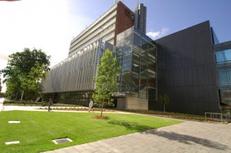
June Griffith Building (F10)
UNSW Sydney, NSW 2052
Access – To discuss training or how your project could benefit from using this microscope, please contact the EMU using the enquiries form or email EMUAdmin@unsw.edu.au
Parent facility
Explore more instruments, facilities & services
Our infrastructure and expertise are accessible to UNSW students and staff, external researchers, government, and industry.


