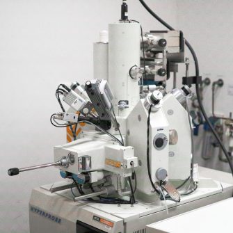
JEOL JSM-7001F FE-SEM
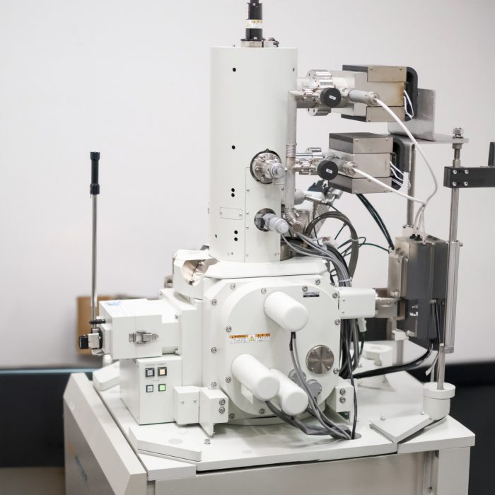
Description
The JEOL JSM-7001F is a high-performance scanning electron microscope (SEM) designed for demanding analytical applications that require high resolution and ease of use. This instrument is equipped with a Schottky field-emission gun, which provides a stable, high-brightness electron source, enabling high-resolution imaging and analysis.
Specifications
-
Schottky field-emission gun
-
SE (Secondary Electron), BSE (Backscattered Electron), EDS (Energy Dispersive X-ray Spectrometer, EBSD (Electron Backscatter Diffraction).
-
0.5-30kV.
-
1.2 nm at 30kV.
-
25x to 1,000,000x.
Publishing Microscopy Data Acquired on the JEOL JXA 8500F
-
-
- Chemical fixation, dehydration, critical point drying
- Mounting in resin
- Staining
- Polishing
- Mounting on stub with adhesive
- Coating
-
- Manufacturer: Joel
- Model: 7001F FE-SEM
- Type: Schottky FEG
-
- Accelerating voltage (kV)
- Detector(s) used for imaging (SE, BSE, EDX)
-
- Detector: EDAX
- Software
- Accelerating voltage (kV)
-
- Adjustments to contrast/brightness
- EDS map filters applied
-
- Scalebar is embedded in the image
Acknowledgement:
“The authors acknowledge the facilities and the scientific and technical assistance of Microscopy Australia at the Electron Microscope Unit (EMU) within the Mark Wainwright Analytical Centre (MWAC) at UNSW Sydney.”
Credit EMU staff: Feel free to mention EMU staff who have assisted you with your work! If staff have been involved with your work beyond basic training and support (e.g., project design, complex data/image processing, independent imaging/analysis, manuscript preparation), it may be appropriate to discuss co-authorship with the relevant staff and your supervisor.
Don’t forget to email the EMU lab manager with a copy of your publication to claim 2 hours of free microscopy time.
-
Applications
- Materials Science
- Life sciences
- Biomaterials
- Solar and battery materials
- Earth Sciences
Capabilities
- Secondary electron imaging
- Backscatter imaging
- Energy dispersive X-ray analysis (EDX)
Instrument location
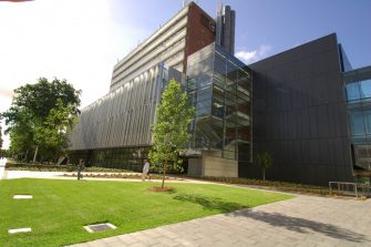
Electron Microscope Unit
B73, Basement
June Griffith Building (F10)
UNSW Sydney, NSW 2052
Access – To discuss training or how your project could benefit from using this microscope, please contact the EMU using the enquiries form or email EMUAdmin@unsw.edu.au
Parent facility
Explore more instruments, facilities & services
Our infrastructure and expertise are accessible to UNSW students and staff, external researchers, government, and industry.
