HeliScan Micro CT-2

High resolution X-ray computed tomography system
Personalise
MWAC Tyree micro-CT lab instrument

Description

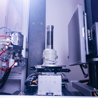
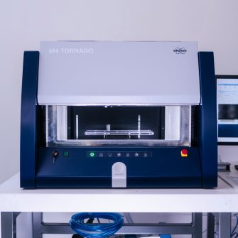
The HeliScan Micro-CT 2 (Mark 1, ThermoFisher) is high resolution X-ray computed tomography in an interlocked lead enclosure system with fluid ports for in situ experiments on small samples. The system allows integration of flow and mechanical cells for semi-dynamic experiment.

The helical trajectory enables longer sample to be scanned in one scan, avoiding post processing for stitching the image. The HeliScan also able to perform volume of interest scanning, by zooming in on selected regions based on the overview image in helical scanning mode.

Specifications

    • X-ray focal spot size at 1.0 μm with ultimate to 0.8 μm. Below table can be used as a guide for sample diameter vs voxel resolution
    Sample Diameter (mm)  Voxel Resolution (µm)
    0.9
     6  2.7 
    10  5.5 
    24  10 
    34.7  15.8 
    62  28 
    90.5  31.6
  • X-ray source: Micro-focus with high flux stability (20-160kV/8W) 
  • X-ray detector: Large area a-Si detector (3,072 × 3,072 pixels) 
  • Sample size (Max. ∅ x h): 220 mm × 220 mm · Max. sample weight: 7.5 kg 
  • Motorization: high precision motorized specimen stage for precision helical trajectory 
  • Scanning trajectory: circular, single helix, double helix

Applications

  • Mining and Subsurface/Petroleum Engineering 
  • Civil Engineering
  • Mechanical Engineering
  • Batteries and Fuel Cells
  • Paleontology
  • Biology
  • Electronics
  • Food science/pharmaceutical industry

Application detail

Mining & subsurface/ Petroleum engineering
Study of minerals in complex matrixes, fluid flows in porous media, 3D pore structure study, in-situ imaging of samples during core flooding (gas/fluid injection), integration of 2D XRF and XRD data into 3D micro-CT data.
Civil engineering
Analysis of internal structure, eg: cracks/voids, analysis of aggregates/impurities inside concrete samples, analysis of structural changes, eg: precipitation in cement/concrete, material enhanced concrete study (eg: fibre reinforced concrete).
Mechanical engineering
Composites (carbon fibres) structure study, in-situ imaging of samples during tensile and compression test, study of 3D prints additive manufacturing (eg: different alloys/polymer).
Batteries & fuel cells
Study of fuel cells materials, the interactions between the different materials and evaluate the quality of the contact between such surfaces. 3D imaging for failure investigation of battery cells.
Paleontology
Analysis of internal structure in bone/fossil, to study bone volume and density, vascularization and trabecular patterns in fragile fossils. Analysis and volumetric measurement of different biological parts, eg: enamel and dentine in tooth.
Biology
Non-destructive technique to study the change in pore geometry at root-soil interface during plant growth. 3D visualization and quantification of root system architecture grown in soil. The technique is also useful to track temporal changes of soil pore architecture due to microbial decomposition of organic matter.
Electronics
PCB’s - 2D/3D imaging for PCB cross-section, and components. It helps identify issues such as board shorts, trace/via connection problems, layout inconsistencies, etching defects, and delamination.
Food science/pharmaceutical industry
3D imaging of emulsion/foam microstructure. By providing detailed 3D imaging, it allows for the study of droplet or bubble size, distribution, and stability, offering insights into formulation quality, texture, and performance for improved product development.

Instrument location

Tyree X-ray micro-CT Facility (Micro-CT)

Room LG22
Lower Ground
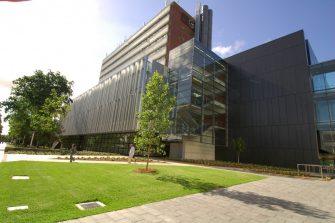
Tyree Energy Technology Building (TETB)

Phone: 02 9385 5554
Emailtyreexray@unsw.edu.au

Amalia Halim

Laboratory Manager
  • Send email envelope icon
    Email
    a.halim@unsw.edu.au

Daniel Morris

Technical Officer
  • Send email envelope icon
    Email
    daniel.morris1@unsw.edu.au

Explore more instruments, facilities & services

Our infrastructure and expertise are accessible to UNSW students and staff, external researchers, government, and industry.