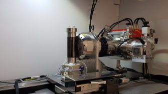
Our workflows are committed to producing the highest quality images and have the capability to analyse very large data sets. Our integrated workflow is aimed at the characterisation of the microstructure of a sample.
The Workflow of a Micro-CT Analysis:
- Preparing the sample to be mounted on the stage
- Acquiring the X-ray CT image
- Reconstructing the 3D tomographic volume
- Identifying phases (segmentation)
- Image registration
There are two main ways the sample is scanned in tomographs. In medical CT machines, the patient (or sample) is held stationary while the X-ray source and detector rotate around the patient. In the micro-CT, the sample needs to be mounted in the stage that rotates either through a double helix pattern or circular pattern while the source and detector are stationary.
In the acquisition process, the detector captures a series of 2D images while the sample rotates. This ensures that the centre of the X-ray beam passes through all parts of the sample. These images are then processed by a computer into a 3D tomogram - this process is known as reconstruction.
After reconstruction, the different parts of the tomogram are identified. In the case of a rock sample, this involves differentiating between the rock matrix and the void space. This process, known as segmentation, may even be extended to differentiating between different minerals in the rock matrix. The reconstructed and segmented tomogram can then be passed through image processing software to create slices or cutaway images and videos. Tomograms can also be analysed to calculate the physical properties of the sample or simulate how fluids would move through them.
Registration is a process of mathematically identifying the exact same space in the two different images. Registration is also used to provide extra contrast (using dry and wet images) and to understand the structural properties in different size sample, imaged at different scales.
Once images have been segmented and registered, it’s possible to combine these identified and labelled volumes for the purpose of morphological and topological classification, including network modelling as well as physical property calculation. The latter—if using the full volume of the image in particular—requires the usage of supercomputing facilities.
A range of solvers for fluid flow, electrical and elastic properties, as well as NMR responses, is available. It is emphasised that some of the latter require significant expert knowledge and applications should be discussed accordingly and usually would be carried out in some form of research collaboration.
The most important thing we offer our clients is quality control, to ensure they get the imagery and/or data that they need. Therefore, we go through a quality control process and assess images for non-resolved features. This assessment may lead to the requirement of integrating other imaging techniques such as SEM (scanning electron microscope) and FIB/SEM by registration. Recently, the elemental mapping from the Itrax Core Scanner has been integrated into the workflow.
Commercial services
Lithium-ion battery
Micro-CT imaging is a powerful tool for lithium-ion batteries failure analysis. Micro-CT allows 3D volumetric observation of the internal parts of the battery to determine the root causes and mechanisms of the failures, such as material degradation, short-circuit (mechanical or electrical), etc
Fibre cement cladding board
3D volumetric evaluation of fibre cement cladding boards damage, detection of flaws such as voids, cracks and particle analysis.
Electronics
Micro-CT can be used for quality control and product defect analysis of various electronics such as electrical circuit board, charger, and mobile phones. We can also provide 2D scan (radiographic) of any electronics for wiring solder bond evaluation.
How research projects use our facility
-
Carbon fibre reinforced polymers (CFRPs) have a well-established role as structural engineering materials, especially in the aerospace industry. High-resolution micro-CT can be used as a non-destructive tool to study the 3D textile architecture of composites at the meso-level without the use of contrast enhancement agents (i.e. uncontaminated). In addition, correct sample preparation (e.g., casting the sample in resin to get cylindrical shape to avoid reconstruction artefact) and advance image segmentation is needed to get the best results from the image. It was found that the Histogram of Oriented Gradients (HOG) gave the best segmentation outcome when the specimen was sized to fit at least two voxels within a fibre width.
The application of these results will assist researchers in better understanding the evolution of microcracks and damage in textile composites while enabling physics-based multiscale modelling approaches to be validated with realistic textile architectures.
-
The Deben CT5000-TEC in-situ micro testing module is a mechanical test stage designed to be used in the confine of X-ray tomography machine or similar apparatus. The module can perform tensile, compression and 3 or 4 points bending test up to a load of 5kN. The cell equipped with a temperature module to conduct test at selected temperature between -20°C and 160°C. Carbon Glass Housing is used for optimal X-ray transparency. Acquisition software which runs on Windows 7 or 10 is used or the control and real-time display of the force-extension curve. The software offers different control mode including load control, displacement control, etc.
-
Micro-CT has become a powerful non-destructive tool to study concrete/composite structure. It enables inspection of the internal structure of material. High-resolution imaging of fibre reinforced concrete allows the optimisation of high-performance concrete by determining spatial fibre distributions and their evolution during flow of the viscous cementitious fluid in the product manufacturing process.
-
Polymer electrolyte membrane fuel cells have a complex 3D structure that requires extensive understanding to improve performances and durability. Micro-CT provides a non-invasive inspection to analyse the interactions between the different materials and evaluate the quality of the contact between such surfaces.
In addition, it enables to study the degradations of the materials via tracking of the crack’s length and depth, alongside thickness monitoring over the lifecycle. Furthermore, it is particularly useful to study compressed areas, as the porosity, tortuosity and permeability can be extracted and modelled from the tomographs.
-
High-resolution micro-CT imaging is a powerful non-destructive tool to study the microstructure of bones and teeth in modern and fossil animals. Parameters such as bone volume, bone density, vascularisation and trabecular patterns can be visualised and measured in fragile fossils using this non-invasive technique. These properties, in turn, provide novel information about the extraordinary lifestyles of long-extinct animals.
-
X-ray microtomography can be used as a non-destructive technique to quantify the changes in pore geometry at the root-soil interface during plant growth. It allows us to perform 3D visualisation and quantification of root system architecture grown in soil. The technique is also useful to track temporal changes of soil pore architecture due to microbial decomposition of organic matter.
-
X-ray microcomputed tomography provides vision into the internal structure of core plugs during core flooding.
The fusion of QEMSCAN and micro-CT data is a powerful tool for 3D mineralogical characterisation of reservoir rocks. These models are the key to understanding complex wetting properties in reservoir rocks that influence multiphase flow. They are also important for the design in situ recovery technologies were a given mineral is targeted for recovery by injecting a reactive agent into the source rock.
-
We present the development and the first trial of a simple X-ray transparent apparatus capable of controlling the sample temperature without influencing the X-ray radiation. The design allows for measurement of axial deformation both in load and displacement control-mode where permeability measurements can be conducted during the experiments. The cell can be used to study the main bedding plane passing through the sample scanned at different confinement loading, at maximum axial stress and confinement unloading.
-
A newly emerging research area is the observation of temporal structural changes when materials are put under loads at high temperature. X-Ray Micro-CT/Neutron Tomography allows the monitoring of the evolution of solid, liquid or gaseous domains in such load cases. A special cell is designed and developed for use in X-Ray micro-CT and Neutron Tomography (in Synchrotron labs). The cells have capabilities of high fluid pressure (100 MPa), high mechanical loads (400 MPa), extreme temperature range (-200 to 700˚C), triaxial and torsion deformation as well as fluid flow.
The prototype X-Ray and Neutron tomography cell design by Dr. Tomasz Blach has been successfully tested in 2018 in the NIST Centre for Neutron Research in Gaithersburg, US and in the Institute Laue Langevin (ILL) in Grenoble, France. Specifications: fluid pressure (up to ca. 100 MPa); hydraulic ram (up to 100 MPa); temperature (up to ca. 300˚C, dual heaters); exchangeable windows (Ti, TiZr, Be, Al); cores (up to 20mm diameter and 100mm long); fluids used (CD4, He, CO2).
-
Recent laboratory studies have shown fines migration induced decrease in rock permeability during CO2 injection. Fines migration is a pore-scale phenomenon, yet previous laboratory studies did not conduct comprehensive pore-scale characterisation. This study utilises integrated pore-scale characterisation techniques to study the phenomenon.
-
High resolution of the images produced by our micro-CT facility allows me to quantitatively analyse the changes in water saturation at different stages of the experiment on the pore scale.
-
For heterogeneous micro-structures the variability in regional structure at the pore scale impacts significantly on the performance of enhanced oil recovery (EOR) processes. We combine high-resolution imaging of heterogeneous porous media with pore-scale rock-typing, physical property calculations and experiments. This provides an integrated micro-scale upscaling workflow calibrated against direct experiments and assists in designing more optimal and realistic EOR and CO2 storage scenarios.
